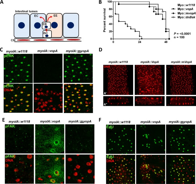
FIG 2.
VopA disrupts epithelial homeostasis in the Drosophila intestine. (A) Schematic depiction of a transverse section of an adult Drosophila midgut. ISC, intestinal stem cell; EC, enterocyte; EB, enteroblast; CM, circular muscle; EE, enteroendocrine cells. Note that the enteroblast in adult Drosophila flies is equivalent to transit-amplifying cells in mammals. ISCs proliferate to generate enteroblasts. The enteroblasts then differentiate to either enterocytes or EEs, which form the gut epithelium in adult Drosophila flies. (B) Life span of adult Drosophila expressing VopA, mVopA, or dominant negative Basket (dnBsk) under myoIA-GAL4. Temperature-sensitive GAL80 (tsGAL80) was also included in each genetic background. Drosophila flies were propagated at 18°C until they were 2-day-old adults, whereupon they were moved to 29°C. Percent survival was recorded at regular intervals. Data are for 100 flies per group. Data are representative of those from experiments undertaken in triplicate. (C) Immunostaining of 7-day-old adult Drosophila midguts of the indicated genotypes in panel B using an antibody against p-ERK. Images were captured by confocal microscopy at a ×40 magnification. (D) Staining of DNA within the distal midgut of Drosophila expressing VopA or mVopA under myoIA-GAL4. (A′) Enface image; (Aʺ) transverse section generated by z-stack analysis of images captured by confocal microscopy. (E) Immunostaining of 7-day-old adult Drosophila posterior midguts dissected from the indicated genotypes from panel B using an antibody against FAK phosphorylated at Tyr397 (p-FAKTyr397). Images captured by confocal microscopy at a ×100 magnification. (F) Detection of proliferating cells in the adult Drosophila posterior midgut by chase analysis. Seven-day-old adult Drosophila flies were fed EdU for 24 h, and cellular EdU incorporation was detected by confocal microscopy at a ×20 magnification. For the images presented in panels C to F, the VopA-mediated phenotype was detected with 100% penetrance, and the images presented are representative of 10 intestinal dissections (n = 10) per group.

