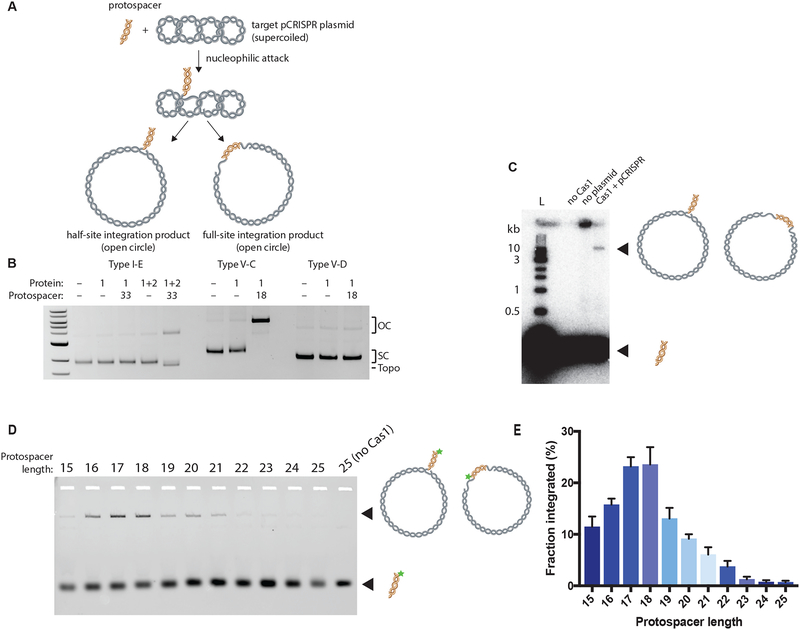
Figure 2. Type V-C Cas1 Integrates DNA Fragments of Expected Length In Vitro.
(A) Schematic of in vitro integration of protospacer into a target supercoiled pCRISPR plasmid. Nucleophilic attack by the protospacer yields two different open-circle products: the half-site and full-site integration products.
(B) Integration assay comparing type I-E, V-C, and V-D systems. A 33-nt protospacer is used for the type I-E system and an 18-nt protospacer for the type V-C and V-D systems. The open-circle integration products (OC), supercoiled target plasmid (SC), and topoisomers (Topo) are indicated.
(C) Integration assay with type V-D Cas1, radiolabeled protospacer, and plasmid target. Integration product and free protospacer are indicated and schematized.
(D) Integration assay with variable length fluorescent protospacers from 15-bp to 25-bp long. Star indicates 6-carboxyfluorescein label.
(E) Quantification of (D) demonstrating the effect of protospacer length on type V-C Cas1 integration. The fraction integrated is calculated as the fraction of the fluorescent protospacer that has been integrated into the target plasmid. Experiments were carried out in triplicate; the bars represent mean values, with error bars depicting standard deviations. See also Figure S1.
See Table S2 for nucleotide sequences.

