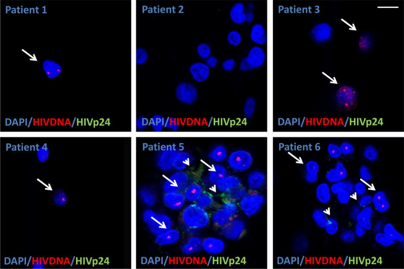
Figure 1.

Detection of HIV Nef DNA and HIV-p24 protein in PBMCs smears obtained from HIV infected individuals with no viral replication detected by at least 5 years. Six representative images of areas with HIV Nef DNA from six different individuals with undetectable replication. Using the technique described, we are able to detect a single copy of HIV integrated DNA (arrows) in non- replicating CD4+ T lymphocytes as determined by ELISA and PCR (>20 copies/ml). To demonstrate that these sequences of integrated DNA are productive, we stained for HIV-p24 (arrowheads). The merged picture shows the colocalization of nuclei (DAPI, blue staining to quantify a total number of cells), HIV DNA (Red nuclear staining), and HIV protein (HIV-p24, green staining). Note that not all cells with inserted DNA produce HIV-p24 protein. Note that in patient 2, we are unable to detect any HIV Nef DNA despite that we analyzed 1.2×10⁷ cells. No positive signal was detected in uninfected individuals with HIV-DNA or HIV-p24 protein. Each picture corresponds just to the positive cells in thousands of negative fields. Most of the circulating cells with HIV Nef DNA were CD4 positive cells. Arrows denote cells with integrated HIV Nef DNA and the arrowhead indicates placed where HIV-p24 is accumulated.
