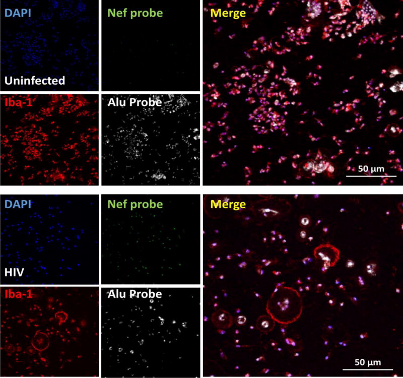
Figure 2.

Detection of HIV Nef DNA, Alu repeats (host DNA), nucleus (DNA) and a macrophage marker (Iba-1) in primary cultures of human macrophages after 2 days post-HIV infection. Using the described technique, we are able to detect the first and maybe the second cycle of HIV replication in these primary cells. Similar results were found using viral reservoirs, latently infected T cells and macrophages. The pictures show the staining of human primary cultures of macrophages for nuclei (DAPI, blue staining to quantify a total number of cells), HIV Nef DNA (green nuclear staining), and a macrophage marker (red staining, Iba-1) in uninfected and HIV infected conditions. Note the perfect colocalization of the Alu repeat and Nef probe as well as DAPI in the HIV infected cultures. This point is essential to demonstrate insertion of HIV DNA into the host DNA.
