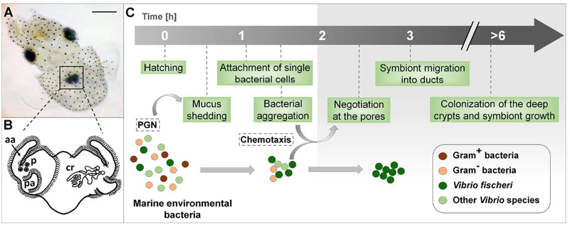
Fig. 1.
The onset of the squid-vibrio symbiosis. (A) A newly hatched Hawaiian bobtail squid, Euprymna scolopes, with its bioluminescent light organ (black square) in the center of the body cavity; scale bar = 500 μm. (B) An illustration of the external (left side) and internal (right side) features of the nascent organ, as viewed ventrally: aa, anterior appendage; cr, crypts; p, pores; pa, posterior appendage. (C) Events in the recruitment of the specific symbiont, Vibrio fischeri, into the organ’s crypts, depicting the stages at which selectivity occurs. The gray shading (right) shows the internal portions of the organ, regions specific to V. fischeri. PGN, peptidoglycan released from bacteria in the ambient seawater.

