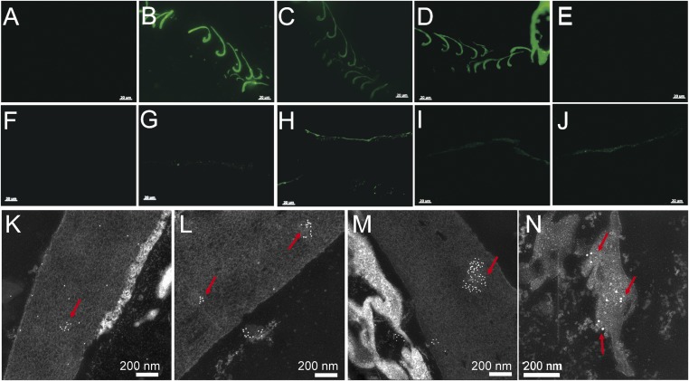
Fig. 4.
In situ IF and IG using four antisera (against extracted feather proteins, Peptide 1, Peptide 2, and broad α-keratin, respectively) on the sample of the pennaceous feathers attached to the right forelimb of Anchiornis and the black flight feather from the chicken G. gallus. All three β-keratin antisera reacted positively with Anchiornis feathers, and in the same pattern as with the chicken feather, but the pan α-keratin antisera reacted only with Anchiornis feather. (A and F) Negative controls, where no primary antibody is applied but all other steps kept identical to test conditions (for additional controls see SI Appendix). (B, G, and K) Exposed to the more broadly cross-reactive antisera against extracted feather proteins. (C, H, and L) Incubated with antisera against feather-specific Peptide 1. (D, I, and M) Exposed to antisera against feather-specific Peptide 2. (E, J, and N) Tested against antisera to the broadly distributed α-keratin. (B–E) IF tests on G. gallus feathers. (G–J) IF tests on Anchiornis feathers. (B–D and G–J) Antibody–antigen (ab-ag) complexes localized to feather tissues from both G. gallus and Anchiornis. (E) Does not show binding to the antiserum against broad α-keratin. (K–N) IG tests on Anchiornis feathers show ab-ag localized to feather tissues. [Scale bars: (A–J), 20 µm; (K–N), 200 nm.]

