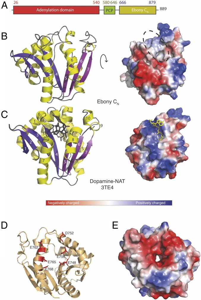
Fig. 2.
Crystal structure of Ebony CN and comparison with D. melanogaster dopamine NAT. (A) Primary architecture of Ebony. (B) Crystal structure of Ebony CN shown as a cartoon (Left) and charge-colored surface (Right); note the splaying in the central β-sheet and the flat, hydrophobic surface in Ebony CN compared with the CoA binding site in the dopamine-NAT homolog. (C) Crystal structure of dopamine-NAT shown as a cartoon (Left) and charge-colored surface (Right); β-strands are in white; helices, in black. (D) Localization of residues in the charged acidic region shown as a cartoon; and (E) overall negative surface charge as visualized in a charge-colored surface.

