Fig. 2.
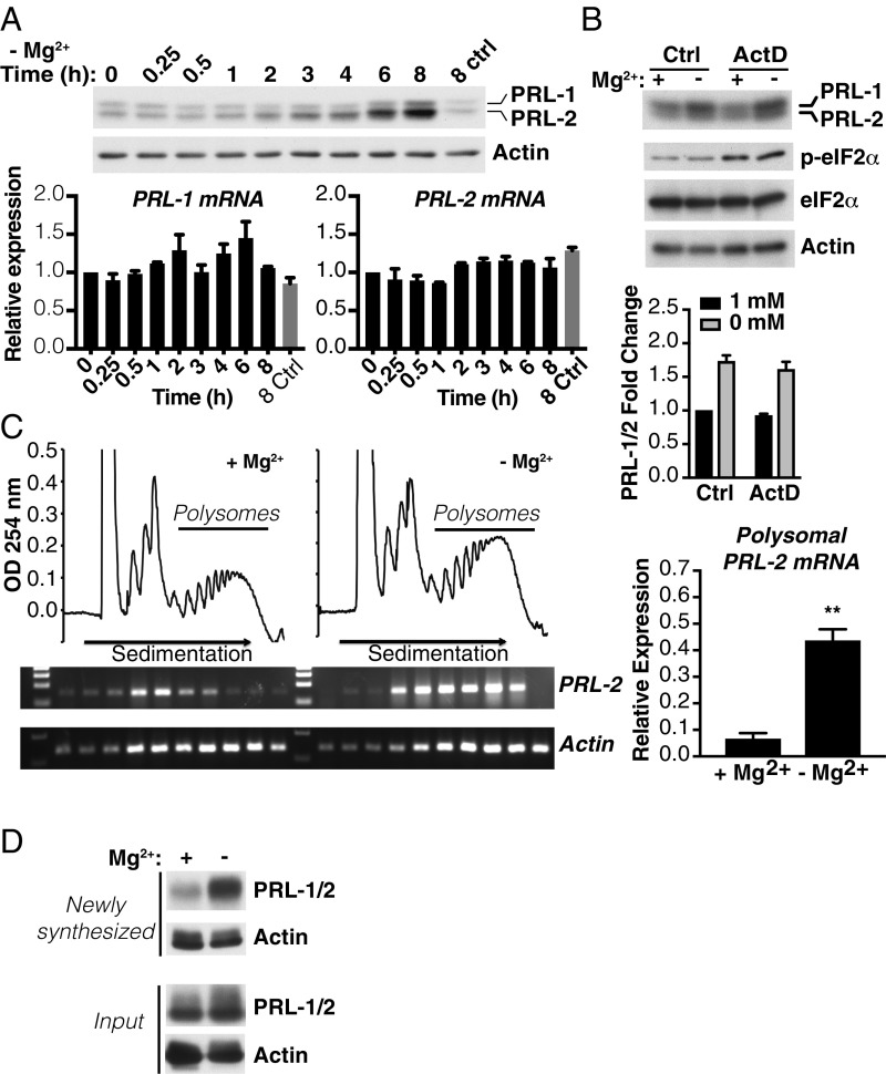
Magnesium depletion promotes PRL mRNA translation. (A) HeLa cells were incubated in the absence of magnesium for the indicated times and analyzed by Western blot (Top) and qPCR (Bottom). The 8 ctrl indicates 8-h control with magnesium. (B) Western blot analysis of HeLa cells incubated for 6 h in either the presence or absence of magnesium with or without actinomycin D (ActD). Quantification is represented as values expressed as fold change relative to 1 mM magnesium control (Ctrl). Data are means ± SEM (n = 3). (C) Polysome profiles of HeLa cells treated for 2 h in either the presence or absence of magnesium. PRL-2 and actin mRNA distribution across the gradient was evaluated in each fraction by semiquantitative RT-PCR and on the polysome fractions by qPCR. Data are means ± SD (n = 3); **P < 0.01 by one-way ANOVA. (D) HeLa cells were metabolically labeled with the methionine analog l-azidohomoalanine for 2 h in either the presence or absence of magnesium. Newly synthesized proteins were covalently bound to biotin, pulled down with streptavidin beads, and analyzed by Western blotting. Input represents total protein extract before biotin pulldown. Representative of three independent experiments.

