Fig. 5.
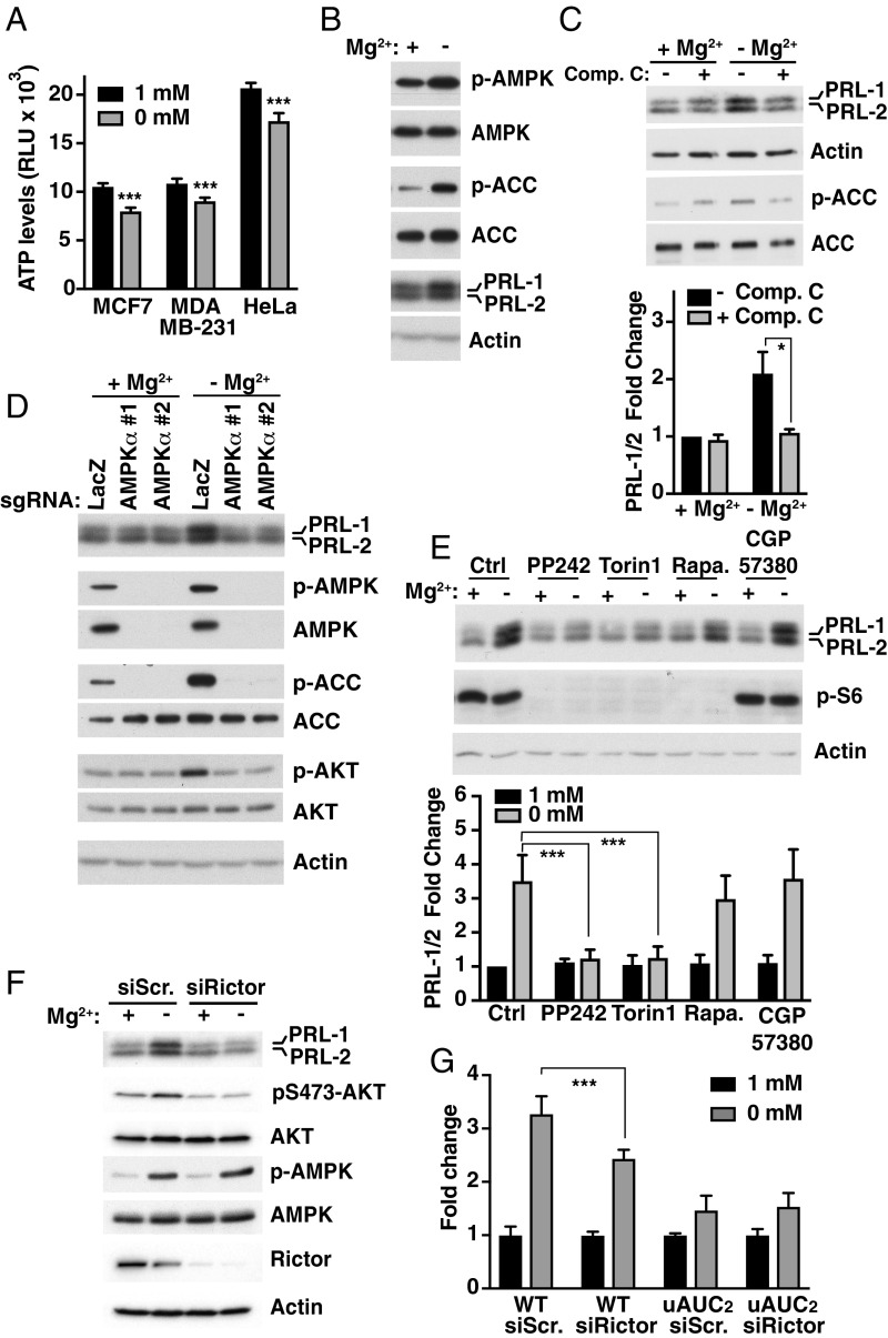
Magnesium regulates PRL expression by an AMPK/mTORC2-dependent pathway. (A) ATP measurements in various cell lines following 2-h magnesium depletion. Data are means data ± SEM (n = 4); ***P < 0.001 by two-way ANOVA. RLU, relative luminescent unit. (B) Western blot analysis of MCF-7 cells incubated for 8 h in either the presence or absence of magnesium. Representative of three independent experiments. (C) MCF-7 cells were incubated for 24 h in the presence or absence of magnesium with or without compound C (Comp. C) and analyzed by Western blot. Quantification is shown as values expressed as fold change relative to 1 mM magnesium without Comp. C. Data are means ± SEM (n = 3); *P < 0.05 by two-way ANOVA. (D) Knockdown of AMPKα using two sgRNAs by the CRISPR-Cas9 system was performed in MCF-7 cells. Targeted cells were incubated for 24 h in the presence or absence of magnesium and analyzed by Western blot. LacZ sgRNA was used as a control. Representative of three independent experiments. (E) MCF-7 cells were incubated for 24 h in the presence or absence of magnesium with or without the indicated inhibitors and analyzed by Western blot. Quantification is shown as values expressed as fold change relative to Ctrl 1 mM magnesium. Data are means ± SEM (n = 3); ***P < 0.001 by two-way ANOVA. Ctrl, vehicle control with DMSO; Rapa, rapamycin. (F) MCF-7 cells were transfected for 30 h with the indicated siRNAs and incubated for 12 h in the presence or absence of magnesium before being analyzed by Western blot. Scr., scramble. (G) HeLa cells were transfected with the indicated constructs and siRNAs followed by luciferase activity measurement. Plot is representative of three independent experiments. Data are means ± SD (n = 4); ***P < 0.001 by two-way ANOVA.

