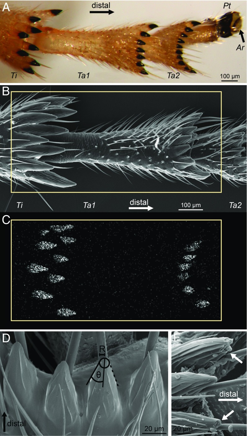
Fig. 1.
Hind leg morphology of P. spumarius froghoppers. (A) Ventral view of distal tibia and tarsus. The dark brown color of the spines indicates strong sclerotization. (B) Scanning electron micrograph of hind leg (ventral view). (C) EDX scan of the same leg as in B, showing the location of zinc (Kα X-ray emission) in the tips of the spines. Rectangle in B shows the area sampled in C. (D) Conical spines on the distal end of the first tarsal segment. (E) Broken spine tips on the first tarsal segment (arrows, ventral view). Ar, arolium; Pt, pretarsus; R, tip radius; Ta1, tarsomere 1; Ta2, tarsomere 2; Ti, tibia.

