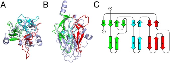
Fig. 4.
The C-terminal domain of PlyB, (A) Ribbon diagram for superimposed C-terminal domains of GNIP1Aa (light blue) and PlyB. Three subdomains of the PlyB β-tripod domain are shown in three different colors: 1 (green), 2 (cyan), and 3 (red). A view along the threefold pseudosymmetry axis. See Fig. 2C for comparison. (B) The same structural alignment with a view perpendicular to A. See Fig. 2D for comparison. (C) Topology diagram for the C-terminal domain of PlyB. The strands are colored and labeled according to their GNIP1Aa counterparts in Fig. 2B.

