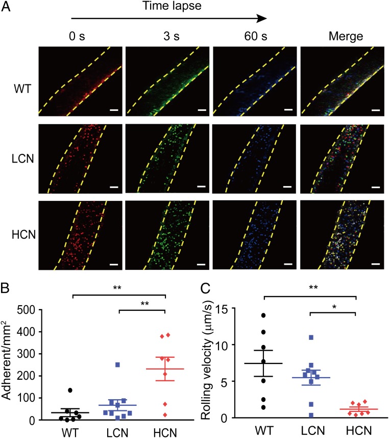
Fig. 2.
Increased gene copy number of DEFA1/DEFA3 exacerbates leukocyte adhesion following sepsis. (A) Representative in vivo time-lapse imaging by two-photon laser scanning microscopy showing leukocyte adhesion in mesenteric vessels in WT mice, mice with LCN of DEFA1/DEFA3, and mice with HCN of DEFA1/DEFA3 at 72 h after CLP. Images are individual frames from a continuous time-lapse movie. Times are presented in seconds. In each group, the first panel shows baseline adhesion in the vessel, the second and third panels show leukocyte adhesion at 3 s and 60 s after the baseline imaging, and the fourth panel shows the merged images. Red indicates leukocyte adhesion at baseline; green indicates leukocyte adhesion at 3 s after baseline; blue indicates leukocyte adhesion at 60 s after the baseline; yellow dashed lines indicate the vessel. (Scale bars, 50 μm.) (B) Quantitative measurements of firmly adhering leukocytes in WT mice, mice with LCN of DEFA1/DEFA3, and mice with HCN of DEFA1/DEFA3 at 72 h after CLP. Firmly adherent leukocytes from at least three fields-of-view per mouse were counted as the number of cells per square millimeter of vascular surface area. **P = 0.002 between WT mice and mice with HCN of DEFA1/DEFA3, **P = 0.007 between mice with LCN of DEFA1/DEFA3 and mice with HCN of DEFA1/DEFA3, one-way ANOVA with Bonferroni posttests. (C) Quantitative measurements of leukocyte rolling velocities in WT mice, mice with LCN of DEFA1/DEFA3, and mice with HCN of DEFA1/DEFA3 at 72 h after CLP. *P = 0.048, **P = 0.005, one-way ANOVA with Bonferroni posttests. In B and C, n = 7 for WT mice with CLP and mice carrying HCN of DEFA1/DEFA3 with CLP, n = 9 for mice carrying LCN of DEFA1/DEFA3 with CLP.

