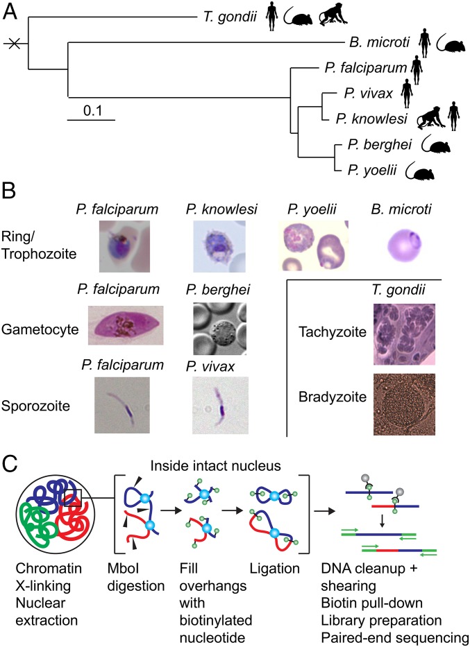
Fig. 1.
Overview of samples and protocol. (A) Phylogenetic tree showing the genetic relationship between the seven different apicomplexan parasites used in this study. Adapted with permission from ref. 61; permission conveyed through Copyright Clearance Center, Inc. (B) Light microscopy images of the various parasites. (C) Schematic representation of the in situ Hi-C protocol.

