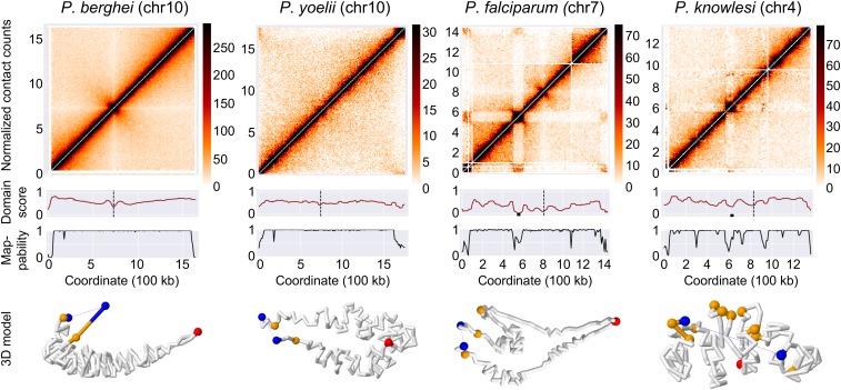
Fig. 4.
Formation of DLS and chromosome loops by var and SICAvar genes. Top row: normalized intrachromosomal contact-count heat maps at 10-kb resolution for representative chromosomes, showing a canonical “X” shape for chromosomes of P. berghei and P. yoelii, and DLS in chromosomes with internal var and SICAvar genes in P. falciparum and P. knowlesi, respectively. Second row: domain score tracks. Dips in the tracks that reach the threshold of a DLS are marked with a black box and centromeres are marked with a black dashed line. Third row: mappability tracks. Bottom row: individual chromosome conformation extracted from the 3D model of the full genome. P. berghei and P. yoelii chromosomes show a folded structure anchored at the centromere, with both chromosome arms arranged in parallel. P. falciparum and P. knowlesi chromosomes show additional folding structures to bring virulence genes in close spatial proximity.

