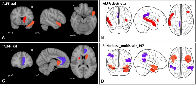
Fig. 5.
Key pathological alterations in schizophrenia suggested by top-most reliable features—elevated (red) and suppressed (blue) changes in regional activity. Panels show top 98th percentile of top regional features. a Higher ALFF in right caudate and right superior temporal pole (aal). b Higher ALFF in lateral aspect of left superior temporal gyrus and horizontal ramus of the right lateral sulcus, and lower ALFF in left posterior-dorsal cingulate gyrus (destrieux). c Higher fALFF in left putamen, right caudate and lower fALFF in right anterior cingulum (aal). d Higher ReHo in left superior temporal pole, right inferior temporal gyrus, and lower ReHo in left inferior parietal lobule and right superior temporal gyrus (basc_multiscale_197)

