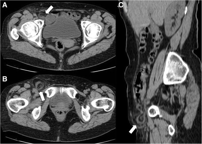
Fig. 10.
Acute epiploic appendagitis in a 59-year-old woman. Axial non-contrast CT images at two different levels (a, b) and sagittal reformatted image (c) show a right inguinal hernia containing an epiploic appendage surrounded by a thin hyperdense rim (arrows). Fat stranding is seen within the hernia sac. Surgical hernia repair and pathological examination confirmed the presence of an epiploic appendagitis incarcerated in inguinal hernia

