
Fig. 13.
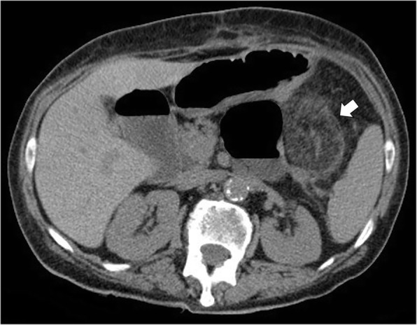
Acute omental infarction in a 67-year-old woman with previous history of total colectomy. Axial non-contrast CT image shows a 6.0 cm heterogeneous fatty ovoid lesion in the left upper quadrant (arrow), associated with inflammatory changes in the nearby fatty tissue