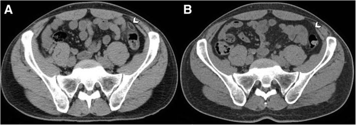
Fig. 7.
Evolution of acute epiploic appendagitis in a 37-year-old man. a Axial non-contrast CT image shows an inflamed fatty ovoid lesion near the sigmoid colon (arrowhead), with hyperattenuating ring sign and nearby fat stranding. b CT scan, performed 1 year later for other reasons, demonstrates complete resolution of imaging findings of epiploic appendagitis (arrowhead)

