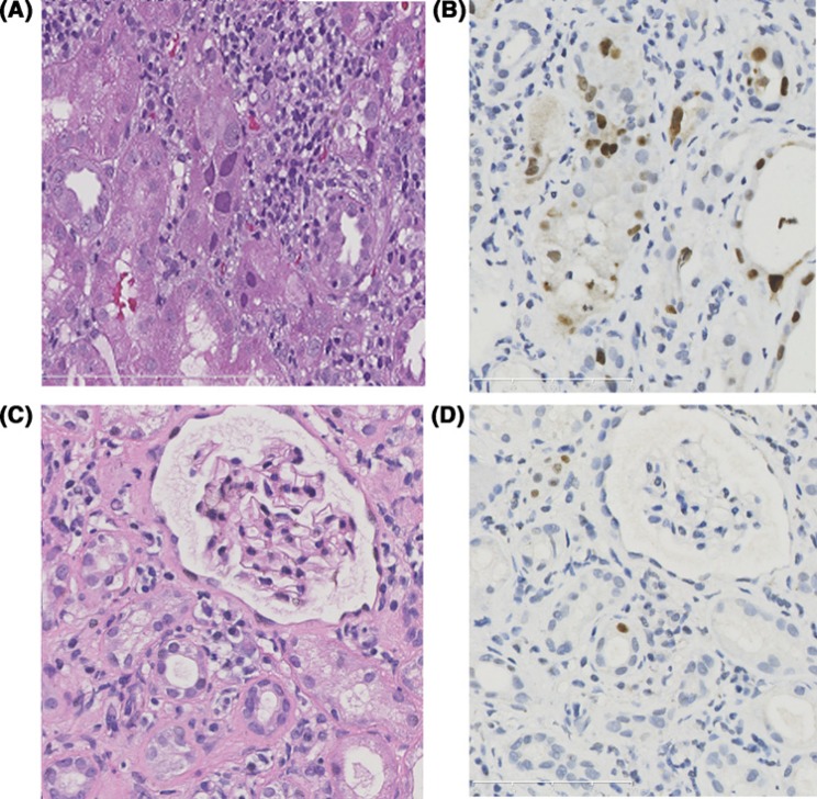
Figure 4. Comparison of representative pathological findings between initial biopsy and repeated biopsy.
BKV cytopathic changes and SV40 large T antigen staining before (A,B) and after (C,D) treatment. Initial biopsy show nuclear enlargement and nuclear inclusions within tubular epithelial cells (A). The SV40 large T antigen staining (B) shows extensive staining in the nuclei in the infected tubules. After switching to low-dose cyclosporine, the viral cytopathic changes become sparse or unrecognizable on Hematoxylin and Eosin staining. SV40 large T antigen staining (D) shows infected nuclei in isolated cells.

