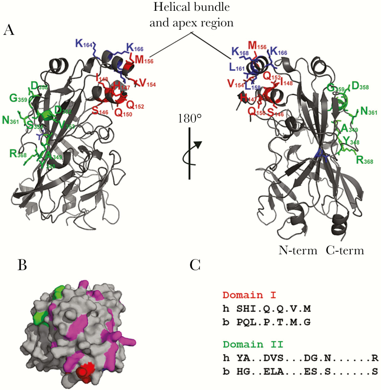
Figure 1.
Human CD36 model. (A) Three-dimensional model based on the crystal structure of lysosomal integral membrane protein (LIMP)-2 [16]. Structure depicted in gray and domains of interest color coded. Domain I in red: SHIQQVM; domain II in green: YADVSDGNR; and other amino acids of interest in blue: T, LLKK. Model generated using PyMol. Helical bundle and apex region marked. (B) The cationic patch on the surface of CD36 viewed from the top. Cationic residues in the area depicted in magenta, whereas flanking domain I (red) and domain II (green) are highlighted. The rest of the structure is shown in gray. (C) Domain I and II alignment between human and bovine CD36 sequences.

