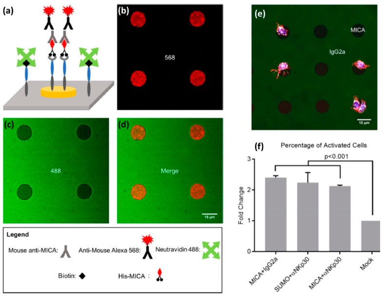
Figure 6.
Site-selective functionalization of a photolithographic pattern with two ligands for NK cell receptors. (a) Orthogonal functionalization of TiO2/Au surface (b) Fluorescent image of Au disks functionalized with His-MICA, and stained with mouse anti-MICA and anti-mouse Alexa 568. (c) Fluorescent image of TiO2 background functionalized with Oregon Green 488-labeled NeutrAvidin. (d) Merging images (c,d) confirm the site-selective functionalization. (e) Primary NK cells stained with Alexa Fluor 555 phalloidin to visualize the cytoskeleton, DAPI for nuclei, and APC-labeled anti-CD107a as a marker for NK cell activation (f) Percentage of CD107a positive cells on various regions of the substrates. Analysis of variance and Tukey’s post hoc test were performed to assess the significant changes in behavior. The results were considered significant for p < 0.05. p-Values are also reported for comparisons of interest. Reprinted with permission from Reference [50].

