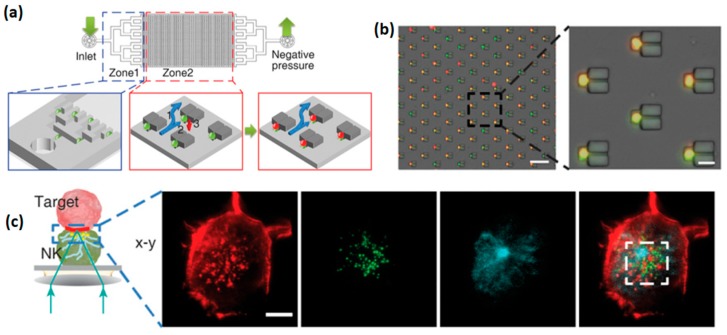
Figure 8.
Microfluidic well arrays with microtraps for NK cells. (a) Scheme of the microfluidic device (b) Merged bright file and fluorescent images after the cell loading into the device; (b) red and green channels correspond to K562 and KHYG-1 cells, respectively. Scale bar, 100 µm (left) and 20 µm (right). (c) Immune synapse imaged by confocal microscopy at the interface of vertically stacked of NK—target cell pair. The cells are fixed, permeabilized, and stained for F-actin (red), perforin (green), and alfa-tubulin (cyan). Scale bars: 5 µm. Reproduced with permission from Reference [65].

