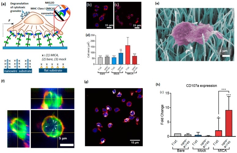
Figure 10.
Nanowire platform for the assessment of NK cell mechanosensing. (a) Cartoon of the experimental setup, and 6 conditions used to assess NK cell mechanical activity. (b,c) NK cells spread on flat MICA and nanowire-MICA surfaces, respectively, (d) Quantification of NK cell spreading on the tested surfaces (** p < 0.001, *** p < 0.0001) (e) Scanning electron microscope image of NK cell applying forces into adjacent nanowires. (f) Z stack of confocal microscopy of NK cells with tagged membrane onto MICA-functionalized, bare, and mock-functionalized nanowires, respectively. Cell membrane (green) and actin (red). The white arrows point onto the projected invaginations of the nanowires in the cell membrane. (g) NK cells stimulated on MICA-functionalized nanowire. Here, CD107a expression was quantified by measuring the fluorescence intensity of the APC-labeled anti-CD107a (in white). (h) Percentage of CD107a positive NK cells on different surfaces normalized surfaces with bare nanoparticles (* p < 0.05, *** p < 0.0001) Copyright Wiley-VCH Verlag GmbH & Co. KGaA. Reproduced with permission [90].

