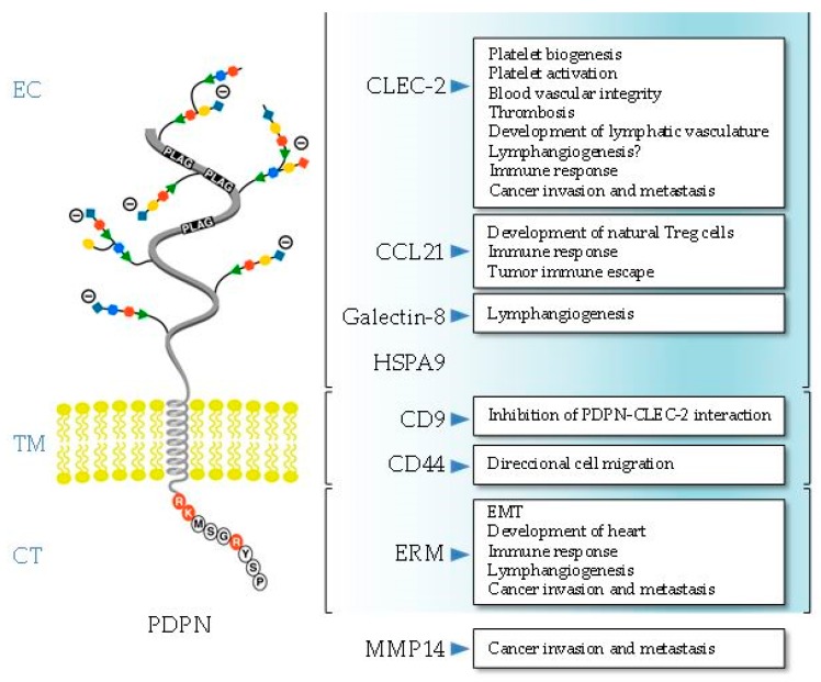
Figure 1.
Schematic representation of the structure of human podoplanin (PDPN) showing the amino-acid sequence of the short cytosolic (CT) domain. The ligands and the biological processes in which the interaction with PDPN is involved are presented. The main PDPN structural domain involved in ligand binding is indicated, except for matrix metalloproteinase 14 (MMP14), which is presently unknown. EC, ectodomain; TM, transmembrane region; CT, cytosolic domain.

