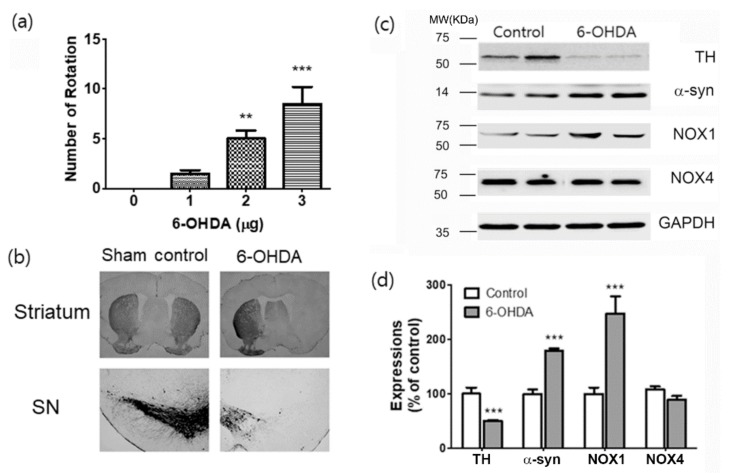
Figure 1.
Evaluations of motor performance, dopaminergic neuronal death, expressions of α -synuclein and NOX1 in the 6-OHDA-induced PD mouse. (a) Total apomorphine (APO)-induced rotation numbers were counted at 4 weeks after 6-OHDA injection. (b) Representative photomicrographs of tyrosine hydroxylase (TH) staining in the mouse striatum and substantia nigra (SN) sections. (c) Results are presented as the mean ± SEM, n = 6. (c) Representative photomicrographs of western blots for α-synuclein (α-syn), NOX1, NOX4, and GAPDH in total lysates of the SN tissue at 4 w after 6-OHDA injection. MW, molecular weight; KDa, KiloDalton. (d) Signal intensities were measured using Quantity One software and are shown as a percentage of control. GAPDH was considered as an internal control. Results are presented as the mean ± SEM, n = 6. *** p < 0.001 vs. control.

