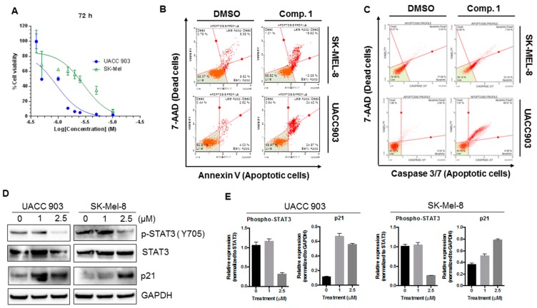
Figure 7.
Compound 1 inhibited the phosphorylation of STAT3, increased the expression of p21, and induced apoptotic cell death in UACC 903 and Sk-Mel-8 melanoma cell lines. (A) Cell viability (MTT) results for compound 1 in two different melanoma cell lines at 72 h. The graphs were obtained by performing non-linear regression analysis using variable slope. Error bars represent mean ± SD. (B,C) Human melanoma cells UACC 903 and SK-Mel-8 were treated with compound 1 (2.5 μM) for 24 h, and an apoptotic assay was performed using the MuseTM Annexin V & Dead Cell (B) and Caspase 3/7 Kit (C) according to the manufacturer’s instructions. (D) Melanoma cells (UACC 903 and SK-Mel-8) cells were treated with the mentioned concentrations of Compound 1. After 24 h, cells were collected and lysed, and the expression of p-STAT3, STAT3, and p21 was analyzed by western blot. GAPDH was used as a loading control; (E) Quantification of protein expression was performed using Image J software, and graphs represent the relative expressions of proteins. The relative expressions were determined using either STAT3 or GAPDH as mentioned.

