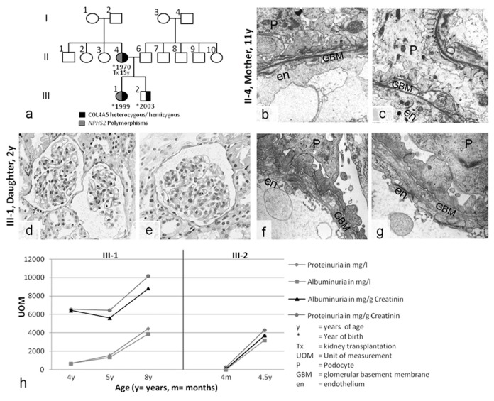
Figure 2.
(a) Pedigree of case 2. (b,c) Kidney biopsy of the mother (11 y) revealing partial GBM thinning, splitting, and laminations in the lamina densa, with plump podocyte (P) foot processes. (d–g) Kidney biopsy of the daughter (3 y). (d,e) Light microscopy showing relatively normal glomerular and tubulointerstitial structures. Electron microscopy uncovered GBM pathology with splitting and thinning, similar to the mother’s nephropathology. (h) The course of disease without therapy in this X-chromosomal COL4A5 genotype was unexpectedly very similar in the heterozygous girl (III-1) and her hemizygous brother (III-2): proteinuria constantly increased into the nephrotic range. Magnification: (b,c) 10,000×, (d,e) 400×, (f,g) 12,500×.

