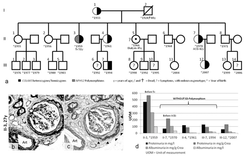
Figure 3.
(a) Pedigree of case 3, with I-1 and family members (II-1, II-2, II-5, II-6, and II-8) living in Poland. Further evaluation was performed on family members living in Vienna (II-3, II-4, II-7, III-7, and III-12). Tx = kidney transplant. (b,c) Kidney biopsy in case II-3. Only a small core of renal tissue could be obtained for light microscopy. This tissue contained only one relatively intact glomerulus and three glomerular scars. The pathology was described as nonspecific and dominated by sclerosis. The glomerulus showed segmental scarring of the capillary loops (open arrowheads) and pronounced periglomerular fibrosis (solid arrowheads). The tubules (T) were dissociated by interstitial fibrosis, and thickening of the tubular basement membranes confirmed advanced atrophy. One small artery (Art) exhibited minimal intimal fibrosis. (d) Proteinuria and albuminuria in family members with and without additional slit diaphragm (SD) polymorphisms. Methenamine silver (b) and PAS staining (c); magnification: 400×.

