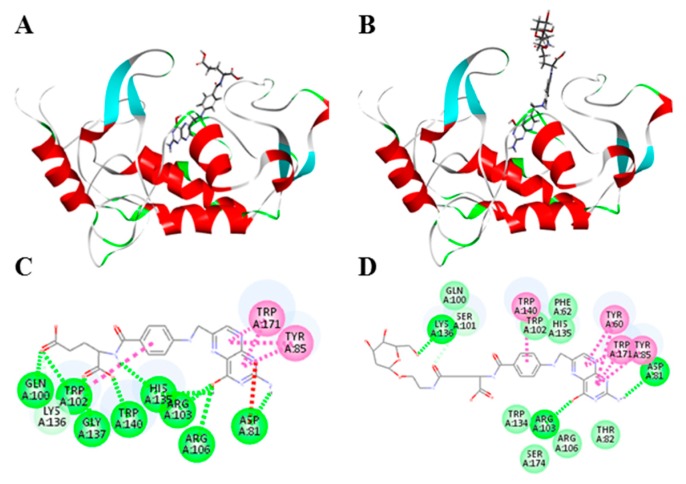
Figure 6.
Docking studies of FA-2-DG with the folate receptor. (A) Folic acid docking into the folate receptor, red color parts represents the alpha helix in the protein structure, light blue color parts represents the beta folding; (B) FA-2-DG docking into the folate receptor, red color parts represents the alpha helix in the protein structure, light blue color parts represents the beta folding; (C) the 2-D diagram of ligand–receptor interactions between folic acid and the folate receptor; (D) 2-D diagram of ligand–receptor interactions between FA-2-DG and the folate receptor.

