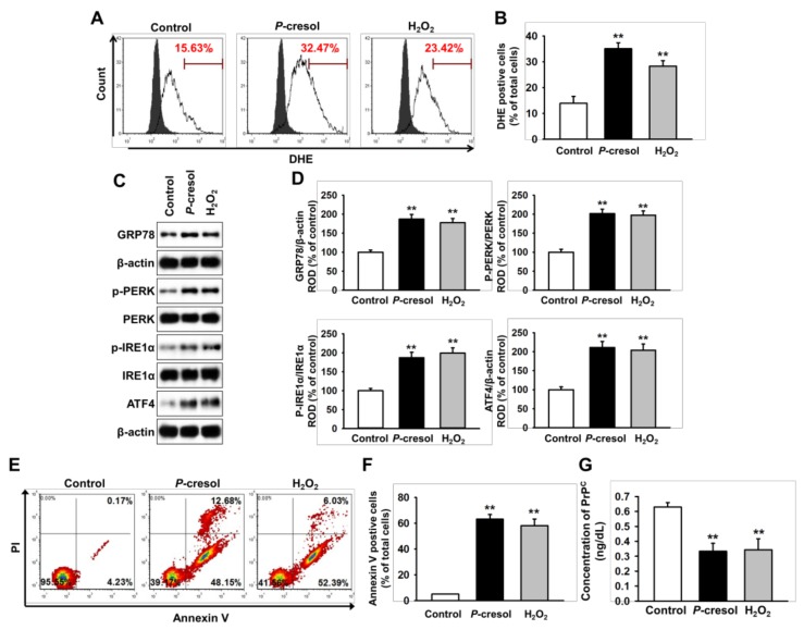
Figure 1.
Uremic toxin-induced apoptosis in SH-SY5Y cells through induction of reactive oxygen species (ROS)-mediated endoplasmic reticulum (ER) stress. (A) Flow cytometry analysis for dihydroethidium (DHE) in SH-SY5Y cells after treatment with P-cresol (500 μM) for 48 h and H2O2 (200 μM) for 4 h (n = 5). The filled and clear histograms represent the cells in the absence and presence of DHE, respectively. (B) Quantification of the percentage of DHE positive cells. (C) Western blot analysis for GRP78, phosphorylation of protein kinase R (PKR)-like endoplasmic reticulum kinase (p-PERK), PERK, phosphorylation of inositol-requiring enzyme 1 α (p-IRE1α), IRE1α, and activating transcription factor 4 (ATF4) in SH-SY5Y cells after treatment with P-cresol and H2O2 (n = 3). (D) The protein levels of (C) were determined by densitometry relative to β-actin. (E) Flow cytometry analysis for PI/Annexin staining in SH-SY5Y cells after treatment with P-cresol and H2O2 (n = 5). (F) Quantification of the percentage of Annexin V positive cells. (G) The concentration of PrPC in SH-SY5Y cells after treatment with P-cresol and H2O2, as assessed by ELISA (n = 5). Statistical analysis: Values represent the mean ± standard error of the mean (SEM). (B) ** p < 0.01 vs. control. (D) ** p < 0.01 vs. control. (F) ** p < 0.01 vs. control.

