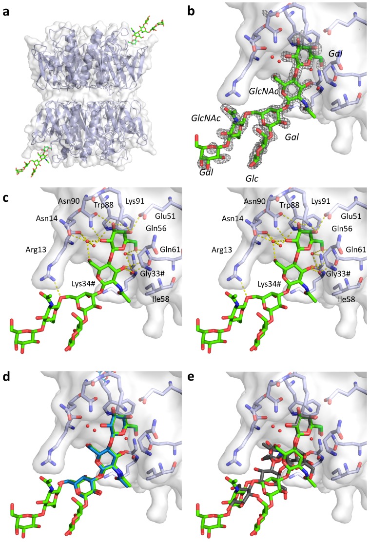
Figure 3.
Structure of pLTB in complex with lacto-N-neohexaose (PDB ID: 6IAL, this work; LNnH shown in stick representation with green carbons). (a) Overview of the asymmetric unit, with two B-pentamers positioned “top-to-top” and two LNnH molecules bound. (b) Close-up view of the ligand binding site, with σA–weighted Fo-Fc electron density map shown in grey mesh contoured at 3.0 σ, generated before placing the ligand. (c) Stereo-image of the binding site, with important residues labeled, H-bonding interactions shown as yellow dotted lines, and selected water molecules depicted as red spheres. Residues from the neighboring subunit are indicated by a hash (#). (d) Close-up view of the LNnH binding site superimposed with LNnT (blue) from structure 2XRS [10]. (e) Close-up view of the LNnH binding site superimposed with GM1 pentasaccharide (grey) from structure 2XRQ [10].

