Figure 3.
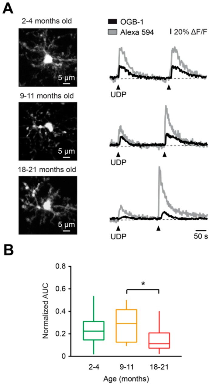
Characterization of UDP-evoked Ca2+ transients in microglia during aging. (A) Representative Ca2+ transients (right) evoked in respective cells (MIP images, left) by two consecutive applications of a solution containing 100 µM UDP and 200 µM Alexa 594 (arrowheads), from pipettes located 30–40 μm away from the cell of interest. (B) Box-and-whisker plot illustrating median (per mouse) normalized AUC (AUCOGB-1 / AUCAF 594) of UDP-induced Ca2+ transients in microglia from 2–4- (green), 9–11- (orange), and 18–21- (red) month-old mice. Normalized AUCs of UDP-induced Ca2+ transients in microglia from 18-21-month-old mice were significantly lower compared to those of 9–11-month-old mice (p < 0.05, Kruskal–Wallis test), but not to those of 2–4-month-old mice (p = 0.13, Kruskal–Wallis test). No significant differences were found between 2–4- and 9–11-month-old mice (p > 0.99, Kruskal–Wallis test; n = 10, 10, and 12 mice for 2–4-, 9–11-, and 18–21-month-old mice, respectively). * p < 0.05 in (B).

