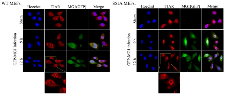
Figure 5.
Formation of stress granules during MG1 infection does not repress viral propagation. WT and S51A MEF cell lines were infected with MG1 virus at MOI = 0.1 for a time course of 12 h as indicated. Immunofluorescence was performed to visualize the stress granules using TIAR anti-rabbit antibody. Cells were imaged by confocal microscopy using 60× water objective. Nuclei (blue) were stained by Hoechst through DAPI filter, GFP-MG1 (green) and TIAR (red) were visualized through FITC and TRITC filters respectively.

