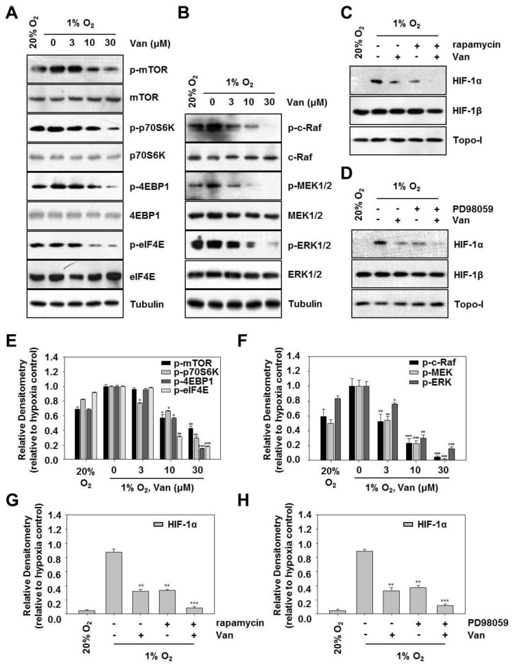
Figure 4.
Vanillic acid (Van) decreased HIF-1α protein synthesis via mTOR/p70S6K/4E-BP1 and Raf/MEK/ERK pathways in HCT116 cells. (A) HCT116 cells were pretreated without or with indicated concentration of vanillic acid (Van), then cultured under normoxic or hypoxic conditions. After 12 h incubation, phospho-mTOR, phospho-p70S6K, phospho-4E-BP1, and phospho-eIF4E were detected by Western blot analysis. The bottom represents the corresponding total protein to show the equal loading of cell lysates. Levels of tubulin were used as a loading control. (E) Data are shown as mean ± SD (n = 3). * p < 0.05, ** p < 0.01, *** p < 0.001, compared with hypoxia control. (B) HCT116 cells were pretreated without or with indicated concentration of vanillic acid (Van), then cultured under normoxic or hypoxic conditions. After 12 h incubation, phospho-c-Raf, phospho-MEK1/2, and phospho-ERK1/2 were detected by Western blot analysis. The bottom represents the corresponding total protein to show the equal loading of cell lysates. Levels of tubulin were used as a loading control. (F) Data are shown as mean ± SD (n = 3). * p < 0.05, ** p < 0.01, *** p < 0.001, compared with hypoxia control. (C,D) HCT116 cells were pretreated without or with indicated concentration of vanillic acid (30 µM), rapamycin (100 nM), and PD98059 (50 µM), then cultured under normoxic or hypoxic conditions. After cells were harvested, the whole-cell lysates for HIF-1β and nuclear extract for HIF-1α was detected by Western blot. Anti-Topo-I antibody was used as a loading control. (G,H) Data were shown as mean ± SD (n = 3). ** p < 0.01, *** p < 0.001, compared with hypoxia control.

