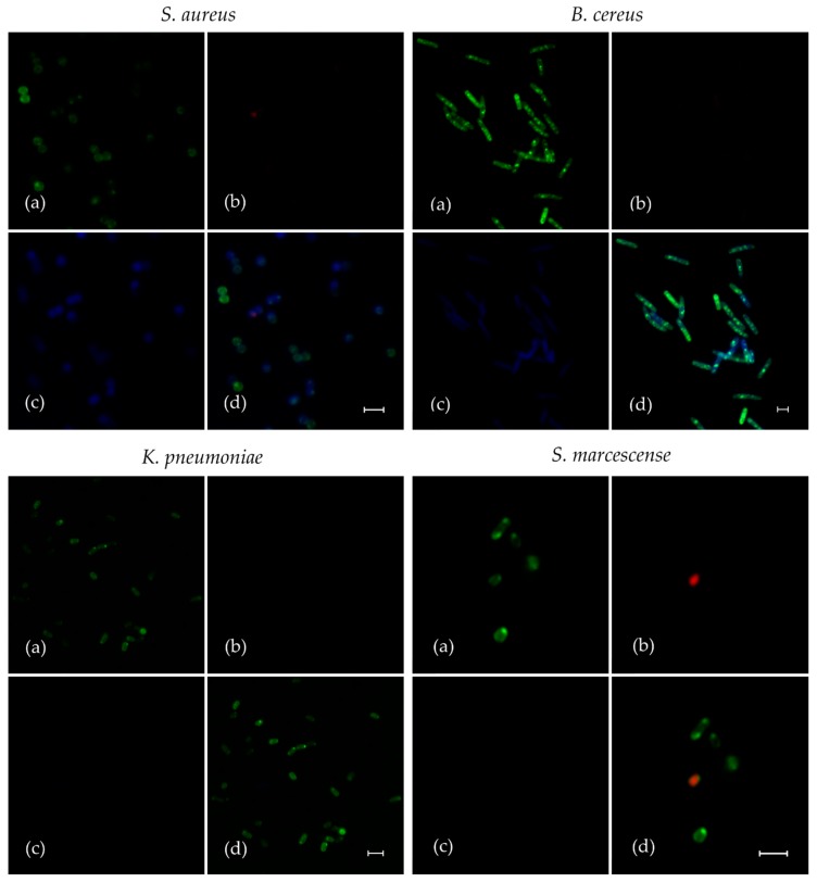
Figure 3.
The analysis of fluorescent F105 analogue (F145) penetration into planktonic bacterial cells. Gram-positive (S. aureus and B. cereus) and Gram-negative (S. marcescens and K. pneumonia) bacteria were grown for 24 h with agitation in MH broth, then washed and resuspended in PBS. F145 was added until the final concentration of 10 μg/mL (close to MIC of F105 for Gram-positive bacteria), and incubation was followed for the next 15 min. Then, the cells were live/dead stained with DioC6/PI (additional 15 min) and analyzed with confocal laser scanning microscopy. Images show the fluorescence of DioC6 (a), PI (b), F145 (c) and the combination of all channels (d). The scale bars indicate 2.5 µm.

