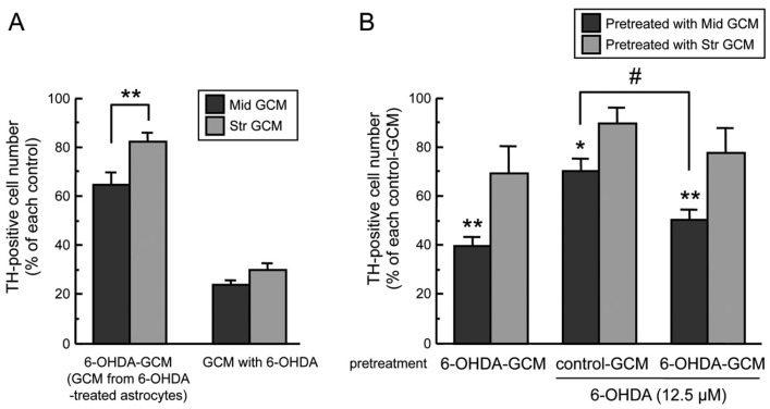
Figure 2.
(A) Regional difference of glia conditioned media (GCM). Cell viability of TH-positive dopaminergic neurons co-incubated with mesencephalic or striatal GCM (control-GCM, 6-OHDA-GCM, GCM with 6-OHDA (100 µM) for 24 h. Each value is mean ± SEM (n = 4) expressed as percentage of each control-GCM group; ** p < 0.01 between indicated two groups. (B) Neuroprotective effect of striatal GCM. Mesencephalic neurons were pre-treated with control-GCM or 6-OHDA-GCM for 24 h, replaced with fresh medium, and then treated with 6-OHDA (12.5 µM) for another 24 h. Data are means ± SEM (n = 3–4) expressed as percentage of TH-positive cell number of vehicle-treated group after each control-GCM pretreatment; * p < 0.05, ** p < 0.01 vs. each control-GCM groups, # p < 0.05 between indicated two groups.

