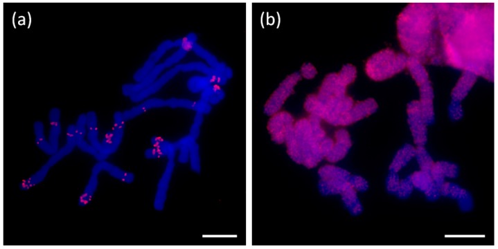
Figure 9.
Fluorescence in situ hybridization with probes containing short tandem motifs. (a) Probe number 11 with the sequence of (TCGGATCGGT)n applied on chromosomes of A. ursinum (b) Probe number 5 with the sequence of (AATCGTTTTCAATTCAATTCATTTCGA)n on chromosomes of A. sativum. The specific probe signals are in red; DAPI staining in blue. The picture (b) exemplifies a dispersed signal pattern obtained for most of the STMs tested (Figure S6). Scale bar, 10 µm.

