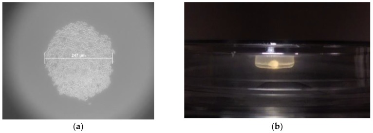
Figure 5.
Phase contrast microscope images of hBM-MSCs mesenspheres. (a) Microscope image of a BM001 hBM-MSCs mesensphere (247 µm largest diameter) grown using the 96-well Perfecta 3D drop plate (100× magnification); (b) Image of a BM001 hBM-MSCs mesensphere (4.6 mm largest diameter) grown using the in-house drop-well device.

