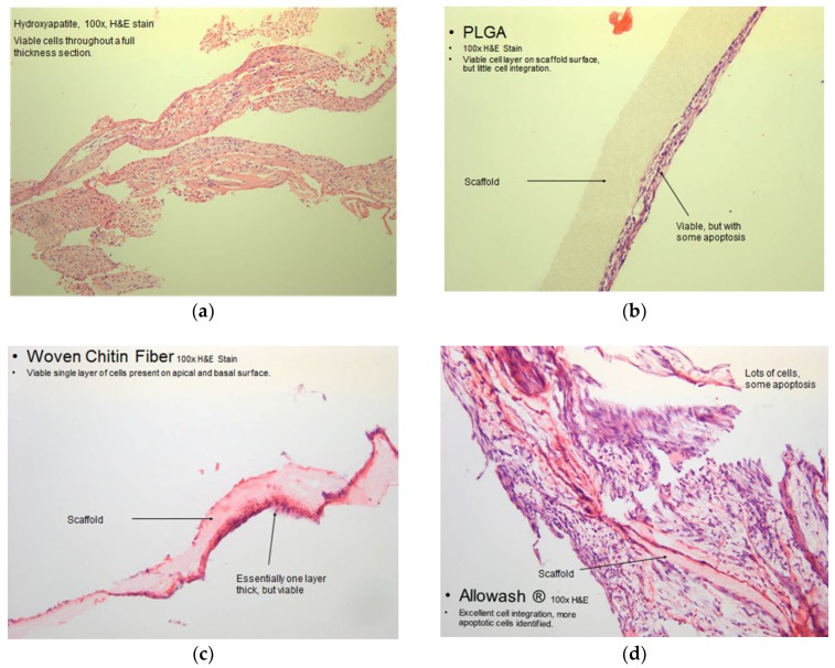
Figure 7.
Microscope image of Haematoxylin and Eosin stained scaffolds following culture with hBM-MSCs. (a) Representative microscope image of a cross-section of hBM-MSCs seeded and cultured on a 60/40 hydroxyapatite scaffold, and stained with Haematoxylin and Eosin (100× magnification). The viable hBM-MSC population can be seen in significant numbers throughout a full thickness section of the scaffold and the cells do not display prominent apoptotic regions.; (b) Representative microscope image of a cross-section of hBM-MSCs seeded and cultured on a PLGA scaffold with 200 µm pores, stained with Haematoxylin and Eosin (100× magnification). The PLGA scaffold did not allow for significant hBM-MSC integration. The MSCs adhered to the outer surfaces of the scaffold and did not migrate far into its interior regions; (c) Representative microscope image of a cross-section of hBM-MSCs seeded and cultured on woven chitin fiber material stained with Haematoxylin and Eosin (100× magnification). Also the woven chitin fiber did not support cell growth to a significant degree, showing a viable single cell layer of hBM-MSC cells on the apical and basal surface; (d) Representative microscope image of a cross-section of hBM-MSCs seeded and cultured on Allowash cancellous bone scaffold stained with Haematoxylin and Eosin (100× magnification). The Allowash cancellous bone scaffold demonstrated excellent cell integration and promoted MSC proliferation.

