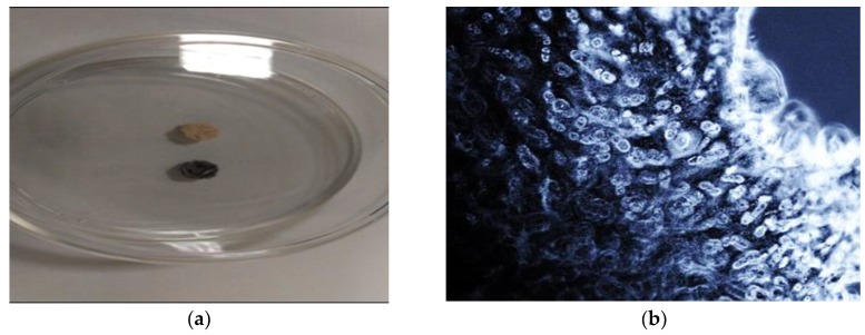
Figure 8.
Images of Tetrazolium salt staining of autograft bone composite following culture with hBM-MSCs. (a) Image of an in-house autograft bone composite disks stained with tetrazolium salt to assess the viability of the cells. A negative control disk, which did not have cells seeded, was used as a control; (b) Inverted light microscopy image of a section from the same scaffold post tetrazolium salt staining (150× magnification).

