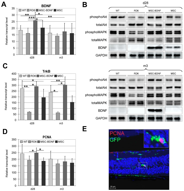
Figure 3.
Long-term follow-up of BDNF production and its biological function at different time points post-intravitreal MSC-BDNF transplantation. BDNF mRNA (A), BDNF, phosphoAkt, totalAkt, phosphoMAPK and totalMAPK protein (B) expression was detected in retinas from eyes treated with MSC-BDNF, and their levels were significantly increased post-transplantation compared to other groups: at 28 days in case of mRNA and protein and at three months in the case of BDNF protein only. We also observed increased TrkB gene expression in retinas at 28 days and three months after MSC-BDNF and MSC alone transplantation compared to rd6 and wild type (WT) (C). The follow-up of retinal cell proliferation at different time points post-intravitreal transplantation in rd6 mice was also performed. Quantitative analysis of proliferating cell nuclear antigen (PCNA) mRNA expression revealed that their levels were significantly increased in retinas from eyes treated with MSC-BDNF at 28 days post transplantation compared with those in eyes treated with the PBS and MSCs alone (D). Double-stained sections for PCNA and GFP (endogenous marker of transplanted MSC) used to visualize and localize proliferating cells revealed the extraordinary PCNA protein concentration in MSC-BDNF transplanted 28 days previously (E). Representative images of the performed analyses are shown. Scale bar: 20 µm. Reference gene used for qRT-PCR analysis was glyceraldehyde 3-phospate dehydrogenase (GAPDH). Mean values ± SD are presented in the diagrams, * p < 0.05, ** p < 0.01, *** p < 0.001 (n = 7/group/time point).

