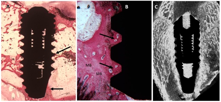
Figure 8.
(A) Trabecular bone was more abundant around the dental implant (arrows). Toluidine blue and acid fuchsin staining 2×. (B) At higher magnification, mature bone (MB) was present around the implant surface and the concavities were completely filled by new mature bone (Arrows). Toluidine blue and acid fuchsin staining 50×. (C) Micro-CT scans along transection planes of the dental implant. Trabecular bone was present in contact with the dental implant and was also more than that in the control implant.

