Figure 2.
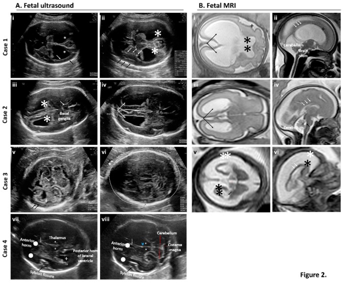
Prenatal examination of brain features by fetal neurosonography (A) and fetal MRI (B) for cases of suspected exposure to ZIKV. (A) Serial and repeat sonographic fetal imaging with neurosonography in the second and third trimester were performed. Case 1: Axial views of the fetal brain at 31 weeks and one day gestational age by best obstetrical estimate (31w1d). There is thinning of the cortex (white arrow) and reduced subarachnoid space (black arrow). Bilateral ventriculomegaly is present (star) (i). At the level of the thalami, (ii) punctiform calcifications are present within the basal ganglia (white arrows) and the subarachnoid space is reduced near the Sylvian fissure (black arrows). The cavum septum pellucidum is present but appears dysplastic (grey arrow). Bilateral ventriculomegaly is present (star). Case 2: Axial views of the fetal brain at 28w5d. Bilateral ventroculomegaly and mild colpocephaly is present (iii, star). The cavum septum pellucidum is present but dysplastic (iv, grey arrow). Bilateral colpocephaly appears asymmetric (iv, arrows). Case 3: Axial views of the fetal brain at 25w5d. There is thalamic hypoplasia (v, white arrow), severe cortical atrophy (v, cross), and a prominent subarachnoid space (v, black arrows). The posterior fossa is normal. (vi) Image is at the level of the transthalamic plane in which the biparietal diameter and head circumference is measured. Microcephaly (defined as head circumference <3SD) is present (dashed line). Case 4: Transthalamic (vii) and transcerebellar (viii) views at 29w3d. No obvious intracranial abnormalities are seen or suspected. Normal structures are labeled. Anterior horns (dots), cavum septum pellucidum (grey arrow), Sylvian fissure (black arrows), thalami (stars), and third ventricle (arrowhead). Cerebellum and cisterna magna (red lines). (B) Fetal MRI with axial views of the transthalamic plane and ventricular system alongside sagittal views of the cortex, cerebellum, brain stem, and corpus callosum revealed abnormalities in three of the four cases. Case 1 (i–ii): Axial and sagittal views at 31w1d. In addition to bilateral ventriculomegaly (i, arrows), there is an abnormal sulcation pattern seen in the Sylvian fissure (i and ii, cross). The leaflets of the cavum septum pellucidum were seen ventrally (not pictured) but not dorsally (i, stars). The corpus callosum was also thin near the splenium (ii, white arrows). The brain stem and cerebellum were normal. Case 2 (iii–iv): Axial and sagittal views at 28w5d. There was mild bilateral ventriculomegaly with a mild colpocephalic appearance (iii, black arrows). The corpus callosum is thin and only partially seen in the area of the splenium (iv, white arrows). The fourth ventricle is not dilated (iv, black arrows) and the brain stem and cerebellum were normal. Case 3 (v–vi): Axial and sagittal views at 25w5d. Severe cortical atrophy is present (v–vi, black stars) with massive enlargement of the subarachnoid space (v–vi, white stars). The lateral ventricles (v, black arrows) are mildly dilated, as is the third ventricle (v, red line). In the midsagittal plane, the corpus callosum is difficult to visualize (vi, white arrows). In real time, the brain matter “jiggled” with fetal movement due to severe atrophy and thinning (supplemental video). An MRI for Case 4 was not performed given the absence of sonographic findings by neurosonography.

