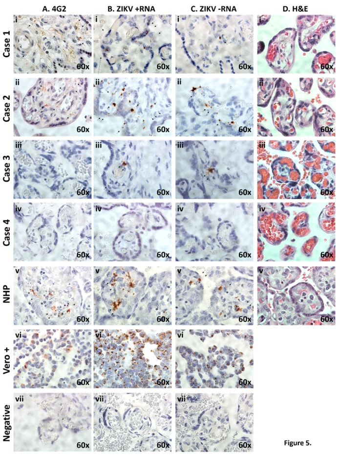
Figure 5.
Histological and IHC examination of placentae reveals evidence of ZIKV infection and active placental replication in congenital Zika syndrome affected cases, but not unaffected. (A) Placental cross-sections were probed using the 4G2 antibody designed against flavivirus envelope protein(s). Diffuse villous and stromal labeling (brown staining, black arrows) were observed in congenital Zika syndrome affected Cases 1 and 2, inconclusive in Case 3, but with no labeling in unaffected Case 4. (B,C) In situ hybridization with oligo probes against plus (+; B) and minus (−; C) ssRNA ZIKV strands were directly assessed with excitation microscopy. Single molecule ssRNA ISH, employing stably amplified ZIKV probes, as previously described [19], was utilized, with summarily positive hybridization (brown labeling, black arrows) in congenital Zika syndrome affected (Cases 1–3) but not in the unaffectedcase (Case 4). In situ ZIKV (+) and (−) strand hybridization was used to distinguish between stable viral strand replicons (plus ssRNA strand, B) and actively replicating (minus strand ssRNA) of the ZIKV genomic replicon, as previously described [19]. (D) H&E-tained sections of placental villus and parenchyma showed no evidence of inflammation in neither congenital Zika syndrome affected nor unaffected cases. No histologic evidence of inflammation was observed with ZIKV infection in any of the human or NHP tissue examined, consistent with previous reports (panel D). In all microscopy experiments shown in panels A–D, comparison with an in vivo infected non-human primate (NHP, Callithrix jacchus) or fixed Vero cells, actively replicating a first passage contemporaneous strain [19] six days post infection, were used as positive controls (vi). To control for non-specific background labeling, parallel placental specimens from Cases 1–4 were imaged after parallel hybridization in the absence of a ZIKV-specific probe (vii).

