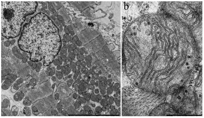
Figure 4.
Electron microscopic images of the ultrastructure of a left ventricular cardiomyocyte from a three-year-old naked mole rat: (a) General view at low magnification. Mitochondrial aggregates in perinuclear area are observed. (b) Ultrastructure of a single mitochondrion at higher magnification.

