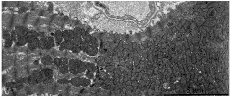
Figure 9.
Electron microscopic image of the ultrastructure of two adjacent left ventricular cardiomyocytes from an 11-year-old naked mole rat. It is visible that the left cardiomyocyte has a normal ultrastructure of mitochondrial apparatus while the right cardiomyocyte contains huge numbers of small mitochondria.

