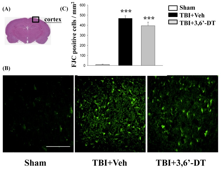
Figure 3.
TBI induces neuron degeneration within the contusion regions, and treatment with 3,6′-DT reduced the number of TBI-induced degenerating neurons. (A) Representative HE-stained coronal brain section from Sham that shows the area of evaluation. (B) Representative photomicrographs showing the presence of fluoro-jade C (FJC)-staining at 24 h in different groups. (C) Quantitative comparison of mean densities of FJC-positive cells in the cortical contusion area at 24 h post-injury. Data are presented as mean ± S.E.M. (n = 5 in each group). *** p < 0.001 compared with the Sham group. Scale bar = 100 µm.

