Figure 10.
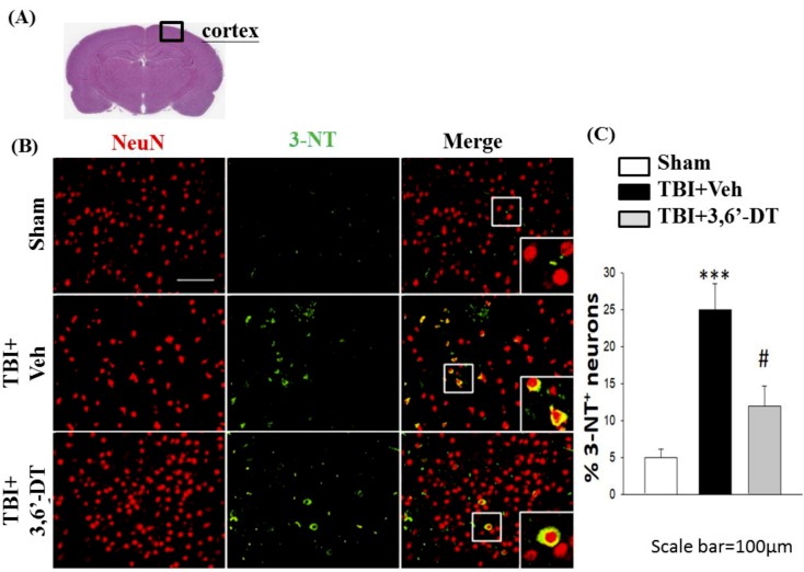
Administration of 3,6′-DT at 5 h post-injury reduced injury-induced tyrosine nitration product in the cortical contusion regions at 24 h. (A) The representative HE-stained coronal section indicates the area used to compare the fluorescent cell observations in different animal treatment groups. (B) The immunofluorescence of 3-nitrotyrosine, 3-NT (tyrosine nitration product mediated by reactive nitrogen species), and NeuN in cortical brain tissue. The 3-NT immunoreactivity is shown in green, and NeuN is shown in red. The yellow color indicates colocalization. (B) Compared to TBI + Veh animals there was a significant decrease in the number of 3-NT/NeuN positive cells in the TBI + 3,6′-DT group. Data are presented as mean ± S.E.M. (n = 5 in each group). *** p < 0.001 compared with the Sham group. # p < 0.05 compared with the TBI + Veh group. Scale bar = 100 µm.

