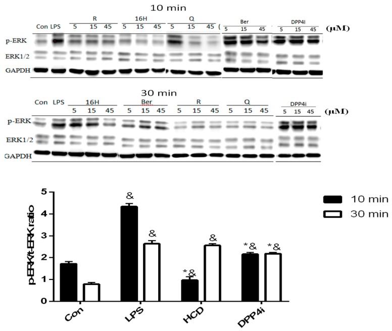
Figure 3.
ERK phosphorylation change after selected natural compounds’ treatment. Myocyte were stimulated by LPS and then treated with, 16-hydroxycleroda-3,13-dien-15,16-olide (HCD & 16H) and sitagliptin (DPP4i) for 10- and 30-min. Ratio of phosphorylated and total ERK levels were detected by Western blotting and normalized with GAPDH. All data were mean ± SD from three independent experiments. * p < 0.05 was marked in the column significantly different to LPS and “&” with DPP4i.

