Figure 2.
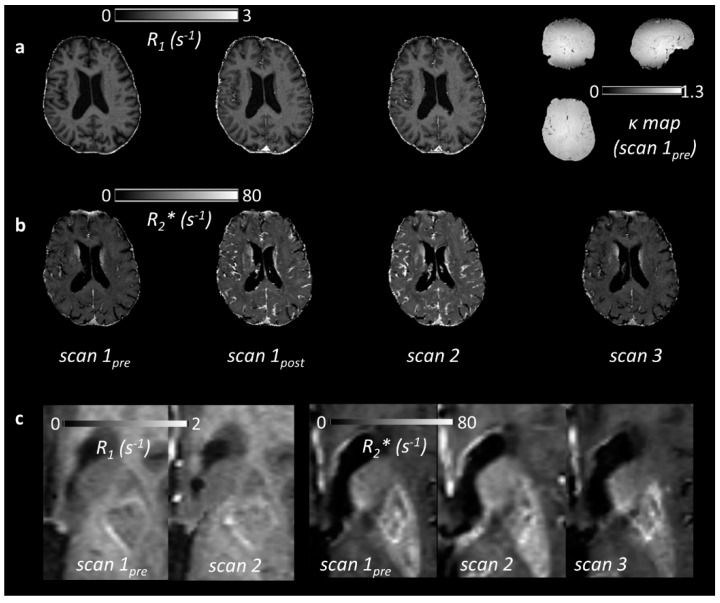
(a) R1 maps for a single patient at three time points: pre-ultrasmall superparamagnetic particles of iron oxide (USPIO) (scan 1pre), immediately post-USPIO (scan 1post), and at 24–30 h post-USPIO (scan 2). The corresponding relative flip angle (i.e., k: the actual flip angle divided by the nominal value) map measured at baseline is shown on the right, illustrating both variation due to B1 inhomogeneity and the (axial) slab excitation profile; (b) R2* maps in the same patient, obtained at the same time points and at 1 month post-USPIO (scan 3); (c) Parametric maps in another patient (79 year old male, first visit 31 days post-infarct in the left basal ganglia). R1 is seen to increase visibly in the stroke lesion following USPIO administration. The corresponding change to R2* is more subtle in the stroke lesion, but a visible increase is seen in the neighbouring background tissue.

